Difference between revisions of "BrainRegionTARGET TSA EYE"
From aHuman Wiki
(Automated page entry using MWPush.pl) |
(Automated page entry using MWPush.pl) |
||
| Line 91: | Line 91: | ||
* [http://usvn.ahuman.org/svn/ahwiki/images/wiki/research/biomodel/vision-subcortical.jpg Vision Components] - see [http://neuroscience.uth.tmc.edu/s3/chapter07.html Reference] | * [http://usvn.ahuman.org/svn/ahwiki/images/wiki/research/biomodel/vision-subcortical.jpg Vision Components] - see [http://neuroscience.uth.tmc.edu/s3/chapter07.html Reference] | ||
| − | <img src="http:// | + | <img src="http://ahuman.org/svn/ahwiki/images/ext/8bc683ef7d36403010f05c36f389cfc9.jpg" alt="unavailable" style="max-width: 100%;"> |
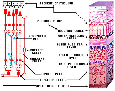
* [http://education.med.nyu.edu/Histology/courseware/modules/eye-and-ear/images/eye-and-ear.21.jpeg Retinal layers] - see [http://education.med.nyu.edu/Histology/courseware/modules/eye-and-ear/eye-and-ear21.html Reference] | * [http://education.med.nyu.edu/Histology/courseware/modules/eye-and-ear/images/eye-and-ear.21.jpeg Retinal layers] - see [http://education.med.nyu.edu/Histology/courseware/modules/eye-and-ear/eye-and-ear21.html Reference] | ||
| − | <img src="http:// | + | <img src="http://ahuman.org/svn/ahwiki/images/ext/6c60ce834c2c3a75de24681cb4a68cdb.jpeg" alt="unavailable" style="max-width: 100%;"> |
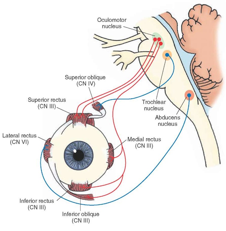
* [http://what-when-how.com/wp-content/uploads/2012/04/tmp15F38_thumb.jpg Extraocular eye muscles] - see [http://what-when-how.com/neuroscience/the-cranial-nerves-organization-of-the-central-nervous-system-part-4 Reference] | * [http://what-when-how.com/wp-content/uploads/2012/04/tmp15F38_thumb.jpg Extraocular eye muscles] - see [http://what-when-how.com/neuroscience/the-cranial-nerves-organization-of-the-central-nervous-system-part-4 Reference] | ||
| − | <img src="http:// | + | <img src="http://ahuman.org/svn/ahwiki/images/ext/28b978f291b0306f8db2e1f3c7bfaf69.jpg" alt="unavailable" style="max-width: 100%;" height=400 width=400> |
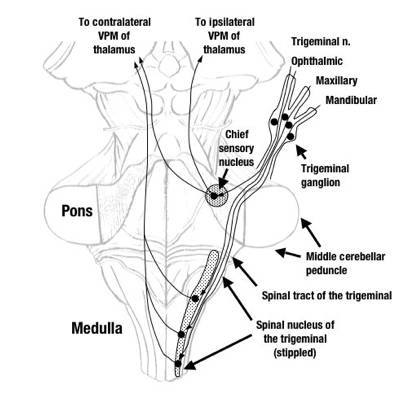
* [http://www.dartmouth.edu/~rswenson/NeuroSci/figures/Figure_16_files/image002.jpg Trigeminal Nuclei] - see [http://www.lookfordiagnosis.com/mesh_info.php?term=Trigeminal+Nucleilang=1 Reference] | * [http://www.dartmouth.edu/~rswenson/NeuroSci/figures/Figure_16_files/image002.jpg Trigeminal Nuclei] - see [http://www.lookfordiagnosis.com/mesh_info.php?term=Trigeminal+Nucleilang=1 Reference] | ||
| − | <img src="http:// | + | <img src="http://ahuman.org/svn/ahwiki/images/ext/8f799fbea59620bf6995c17e1f8b6257.jpg" alt="unavailable" style="max-width: 100%;"> |
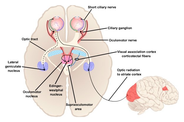
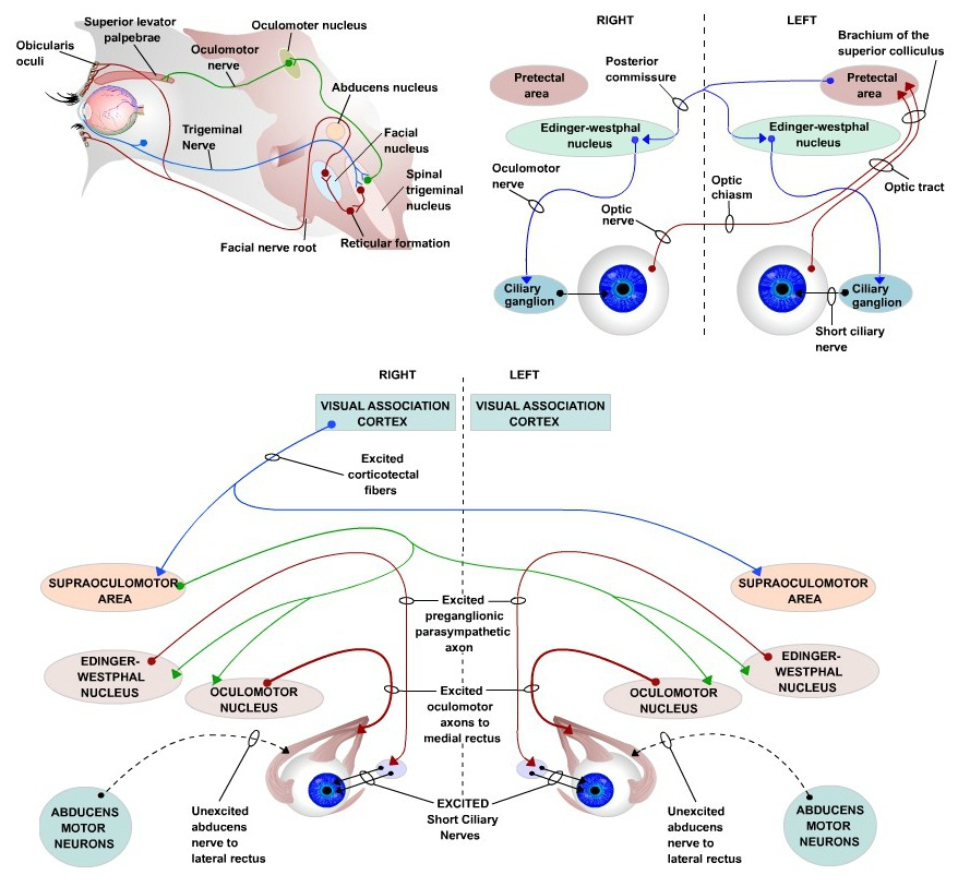
* [http://usvn.ahuman.org/svn/ahwiki/images/wiki/research/biomodel/oculomotor.jpg Eye blink reflex and pupillary light reflex] - see [http://neuroscience.uth.tmc.edu/s3/chapter07.html Reference] | * [http://usvn.ahuman.org/svn/ahwiki/images/wiki/research/biomodel/oculomotor.jpg Eye blink reflex and pupillary light reflex] - see [http://neuroscience.uth.tmc.edu/s3/chapter07.html Reference] | ||
| − | <img src="http:// | + | <img src="http://ahuman.org/svn/ahwiki/images/ext/5fa88e17ecb33cc14ac9fd42f88cea9f.jpg" alt="unavailable" style="max-width: 100%;"> |
* [http://www.frontiersin.org/files/Articles/31465/fnint-06-00094-r2/image_m/fnint-06-00094-g002.jpg Effect of light on pupil diameter] - see [http://journal.frontiersin.org/article/10.3389/fnint.2012.00094/full Reference] | * [http://www.frontiersin.org/files/Articles/31465/fnint-06-00094-r2/image_m/fnint-06-00094-g002.jpg Effect of light on pupil diameter] - see [http://journal.frontiersin.org/article/10.3389/fnint.2012.00094/full Reference] | ||
| − | <img src="http:// | + | <img src="http://ahuman.org/svn/ahwiki/images/ext/1cde10342c1a8c5667b42d9df87c4f50.jpg" alt="unavailable" style="max-width: 100%;"> |
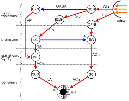
* [http://www.nature.com/pr/journal/v71/n3/images/pr201138f1.gif Sympathetic and parasympathetic eye pathways] - see [http://www.nature.com/pr/journal/v71/n3/fig_tab/pr201138f1.html Reference] | * [http://www.nature.com/pr/journal/v71/n3/images/pr201138f1.gif Sympathetic and parasympathetic eye pathways] - see [http://www.nature.com/pr/journal/v71/n3/fig_tab/pr201138f1.html Reference] | ||
| − | <img src="http:// | + | <img src="http://ahuman.org/svn/ahwiki/images/ext/6f9c7340846328c7a7cc1ccfc7300031.gif" alt="unavailable" style="max-width: 100%;" height=400 width=400> |
* [http://www.neurophysiology.ws/images/fig-4.gif Autonomic nervous system] - see [http://www.neurophysiology.ws/autonomicns.htm Reference] | * [http://www.neurophysiology.ws/images/fig-4.gif Autonomic nervous system] - see [http://www.neurophysiology.ws/autonomicns.htm Reference] | ||
| − | <img src="http:// | + | <img src="http://ahuman.org/svn/ahwiki/images/ext/afc740335d4c9a62e52cca60cc819e98.gif" alt="unavailable" style="max-width: 100%;"> |
Revision as of 12:53, 15 June 2016
Eye
@@Home -> BiologicalLifeResearch -> BrainAreaTSA -> BrainRegionTARGET_TSA_EYE
This page covers biological details of component Eye. Region is part of aHuman target integrated biological model.
- Top-down path to region: Target -> Human Sensors (aHumanSensors) -> Eye (TARGET.TSA.EYE) (see Mind Maps)
- Type: sensor
- Brain area: Sensor Area
- Role: targetsensor
- Function: Light and Color Perception Sensor
(generated)
Components
(generated)
Component items:
- Eye sensory data (eye-sensory)
- Eye - rods and cones (eye-edge-output -> LPC.PN.SG.RT): after on-center/off-center edge extraction and zipping 100:1
- Eye - somatic receptors (eye-somatic -> LPC.PN.SG.TRG): eye -> semilunar ganglion/ophthalmic -> PTR
- Eye - visceral receptors (eye-visceral -> LPC.MM.MSTR): eye -> semilunar ganglion/ophthalmic -> STR, proprioception
- Eye rotation motor muscles (eye-rotation-motor)
- Eye - inferior oblique muscle (LPC.MM.OCN -> eye-muscle-rotate-major-inferior-oblique): extorsion, elevation, abduction
- Eye - inferior rectus muscle (LPC.MM.OCN -> eye-muscle-rotate-major-inferior-rectus): depression and adduction
- Eye - medial rectus muscle (LPC.MM.OCN -> eye-muscle-rotate-major-medial-rectus): adducts the eyeball
- Eye - superior rectus muscle (LPC.MM.OCN -> eye-muscle-rotate-major-superior-rectus): elevates, adducts, and rotates medially the eye
- Eye - levator palpebrae superioris muscle (LPC.MM.OCN -> eye-muscle-rotate-major-levator-palpebrae): retracts/elevates eyelid
- Eye - superior oblique muscle (LPC.MM.TRH -> eye-muscle-rotate-minor): superior oblique, rotation in a vertical plane - looking down and up, rotation in the plane of the face
- Eye - lateral rectus muscle (LPC.HT.ABD -> eye-muscle-rotate-vestibular): lateral rectus, quick turn to the right -> compensatory reflex turning of two eyes to the left
- Eye pupil and accomodation motor muscles (eye-pupil-motor)
- Eye - ciliary muscle - sympathetic (LAC.PN.SYM.SCG -> eye-muscle-curvature-sympathetic): to ciliary muscle to increase eye accomodation
- Eye - ciliary muscle - parasympathetic (LAC.PN.PSYM.CLG -> eye-muscle-curvature-parasympathetic): to ciliary muscle to inhibit eye accomodation
- Eye - iris sphincter muscle (LAC.PN.PSYM.EPG -> eye-muscle-pupil-close): to sphincter pupillae to close pupil
- Eye - iris dilator muscle (LAC.PN.SYM.SCG -> eye-muscle-pupil-open): to dilator pupillae to open pupil
- Eyelid motor muscles (eyelid-motor)
- Eye - orbicularis oculi muscle:temporal (LPC.HT.FCM -> eye-muscle-close-conscious-orbital): orbital portion, contraction of the orbital to reduce glare
- Eye - orbicularis oculi muscle:zygomatic (LPC.HT.FCM -> eye-muscle-close-conscious-lacrimal): lacrimal part (tensor tarsi), conscious muscle to close eye; draws the eyelids and the ends of the lacrimal canals medialward and compresses them against the surface of the globe of the eye; compresses the lacrimal sac
- Eye - orbicularis oculi muscle:palpebrale (LPC.HT.FCM -> eye-muscle-close-involuntary-palpebrale): palpebral portion, acts involuntarily, closing the lids gently; involuntary muscle to close eye
- Eye - superior tarsal muscle (LAC.PN.SYM.SCG -> eye-muscle-superior-tarsal): raise the upper eyelid
- Eye - inferior tarsal muscle (LAC.PN.SYM.SCG -> eye-muscle-inferior-tarsal): lower lid retraction
- Eye - orbitalis muscle (LAC.PN.SYM.SCG -> eye-orbitalis-muscle): protrusion of eyeball
- Eye glands (eye-glands)
- Eye - lacrimal gland (LAC.PN.PSYM.SPG -> eye-tears-gland): tears
- Eye - lacrimal gland and conjunctiva receptors (eye-lacrimal-gland-conjunctiva-receptors -> LPC.PN.SG.TRG): tears
Connectivity
(generated)
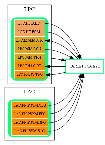
Inbound Region Connections:
| Source Area | Source Region | Source Name | Type | Reference |
| LAC | LAC.PN.PSYM.CLG | Ciliary Ganglion | eye-muscle-curvature-parasympathetic | Autonomic nervous system (CLG -> EYE) |
| LAC.PN.PSYM.EPG | Episcleral Ganglion | eye-muscle-pupil-close | Autonomic nervous system (EPG -> EYE) | |
| LAC.PN.PSYM.SPG | Sphenopalatine Ganglion | eye-tears-gland | (unknown reference) | |
| LAC.PN.SYM.SCG | Superior Cervical Ganglion | eye-muscle-curvature-sympathetic | Sympathetic and parasympathetic eye pathways (SCG -> EYE) | |
| LPC | LPC.HT.ABD | Abducens Nucleus | eye-muscle-rotate-vestibular | Eye blink reflex and pupillary light reflex (Abducens -> EYE) |
| LPC.HT.FCM | Facial Motor Nucleus | eye-muscle-close-conscious-lacrimal | (unknown reference) | |
| LPC.MM.OCN | Oculomotor Nucleus | eye-muscle-rotate-major-inferior-oblique | Eye blink reflex and pupillary light reflex (Oculomotor -> EYE) | |
| LPC.MM.TRH | Trochlear Nucleus | eye-muscle-rotate-minor | Extraocular eye muscles (TRC -> EYE) |
Outbound Region Connections:
| Target Area | Target Region | Target Name | Type | Reference |
| LPC | LPC.MM.MSTR | Mesencephalic Trigeminal Nucleus | eye-visceral | (unknown reference) |
| LPC.PN.SG.RT | Retinal Ganglion | eye-edge-output | Vision Components (EYE -> RT) | |
| LPC.PN.SG.TRG | Trigeminal Ganglion | eye-lacrimal-gland-conjunctiva-receptors | (unknown reference) |
Thirdparty Circuits
(generated)
- Vision Components - see Reference
- Retinal layers - see Reference

- Trigeminal Nuclei - see Reference

References
(generated)
- http://neuroscience.uth.tmc.edu/s3/chapter07.html
- http://education.med.nyu.edu/Histology/courseware/modules/eye-and-ear/eye-and-ear21.html
- http://what-when-how.com/neuroscience/the-cranial-nerves-organization-of-the-central-nervous-system-part-4
- http://www.lookfordiagnosis.com/mesh_info.php?term=Trigeminal+Nucleilang=1
- http://journal.frontiersin.org/article/10.3389/fnint.2012.00094/full
- http://www.nature.com/pr/journal/v71/n3/fig_tab/pr201138f1.html
- http://www.neurophysiology.ws/autonomicns.htm