Difference between revisions of "BrainRegionLPC PN SG RT"
From aHuman Wiki
(Automated page entry using MWPush.pl) |
(Automated page entry using MWPush.pl) |
||
| Line 1: | Line 1: | ||
| − | |||
<pre style="color: green">Retinal Ganglion</pre> | <pre style="color: green">Retinal Ganglion</pre> | ||
@@[[Home]] -> [[BiologicalLifeResearch]] -> [[BrainAreaLPC]] -> [[BrainRegionLPC_PN_SG_RT]] | @@[[Home]] -> [[BiologicalLifeResearch]] -> [[BrainAreaLPC]] -> [[BrainRegionLPC_PN_SG_RT]] | ||
| Line 47: | Line 46: | ||
* [http://education.med.nyu.edu/Histology/courseware/modules/eye-and-ear/images/eye-and-ear.21.jpeg Retinal layers] - see [http://education.med.nyu.edu/Histology/courseware/modules/eye-and-ear/eye-and-ear21.html Reference] | * [http://education.med.nyu.edu/Histology/courseware/modules/eye-and-ear/images/eye-and-ear.21.jpeg Retinal layers] - see [http://education.med.nyu.edu/Histology/courseware/modules/eye-and-ear/eye-and-ear21.html Reference] | ||
| − | map: GANGLION=[[BrainRegionLPC_FD_RT|Retinal Nucleus,LPC.FD.RT]] | + | map: |
| − | <img src="http://ahuman.org/svn/ahwiki/images/ext/6c60ce834c2c3a75de24681cb4a68cdb.jpeg" alt="unavailable" style="max-width: 100%;"> | + | GANGLION=[[BrainRegionLPC_FD_RT|Retinal Nucleus,LPC.FD.RT]] |
| + | INNER=[[BrainRegionLPC_PN_SG_RT|Retinal Ganglion,LPC.PN.SG.RT]] | ||
| + | RODSCONES=[[BrainRegionTARGET_TSA_EYE|Eye,TARGET.TSA.EYE]] | ||
| + | <img src="http://ahuman.org/svn/ahwiki/images/ext/6c60ce834c2c3a75de24681cb4a68cdb.jpeg" alt=alt="unavailable" style="max-width: 100%;"> | ||
* [http://usvn.ahuman.org/svn/ahwiki/images/wiki/research/biomodel/vision-subcortical.jpg Vision Components] - see [http://neuroscience.uth.tmc.edu/s3/chapter07.html Reference] | * [http://usvn.ahuman.org/svn/ahwiki/images/wiki/research/biomodel/vision-subcortical.jpg Vision Components] - see [http://neuroscience.uth.tmc.edu/s3/chapter07.html Reference] | ||
| − | map: CG=[[BrainRegionLAC_PN_PSYM_CLG|Ciliary Ganglion,LAC.PN.PSYM.CLG]] | + | map: |
| − | <img src="http://ahuman.org/svn/ahwiki/images/ext/8bc683ef7d36403010f05c36f389cfc9.jpg" alt="unavailable" style="max-width: 100%;"> | + | CG=[[BrainRegionLAC_PN_PSYM_CLG|Ciliary Ganglion,LAC.PN.PSYM.CLG]] |
| + | EWN=[[BrainRegionLAC_MM_EWN|Edinger-Westphal Nucleus,LAC.MM.EWN]] | ||
| + | EYE=[[BrainRegionTARGET_TSA_EYE|Eye,TARGET.TSA.EYE]] | ||
| + | LGN=[[BrainRegionNSA_FD_LGN|Lateral Geniculate Nucleus,NSA.FD.LGN]] | ||
| + | OCN=[[BrainRegionLPC_MM_OCN|Oculomotor Nucleus,LPC.MM.OCN]] | ||
| + | RT=[[BrainRegionLPC_PN_SG_RT|Retinal Ganglion,LPC.PN.SG.RT]] | ||
| + | SOA=Supraoculomotor Area,VBA.MM.SOA (see [[BrainCenterMM_REF|Reflexes Nuclei of Midbrain Mesencephalon]]) | ||
| + | STN=[[BrainRegionLPC_HL_TGS_STR|Spinal Trigeminal Nucleus,LPC.HL.TGS.STR]] | ||
| + | VAC=Posterior Eye Field,NPE.NC.IPS.PEF (see [[BrainCenterNSA_NC_IPS|Intraparietal Sulcus]]) | ||
| + | VC=[[BrainCenterNC_LOBE_OCC|Occipital Lobe,NC.LOBE.OCC]] | ||
| + | <img src="http://ahuman.org/svn/ahwiki/images/ext/8bc683ef7d36403010f05c36f389cfc9.jpg" alt=alt="unavailable" style="max-width: 100%;"> | ||
Revision as of 18:59, 28 November 2018
Retinal Ganglion
@@Home -> BiologicalLifeResearch -> BrainAreaLPC -> BrainRegionLPC_PN_SG_RT
This page covers biological details of component Retinal Ganglion. Region is part of aHuman target integrated biological model.
- Top-down path to region: Peripheral Nervous System -> Sensory Ganglia (PNS.SG) -> Sensory Exteroception Ganglia (PN.SG.SE) -> Retinal Ganglion (LPC.PN.SG.RT) (see Mind Maps)
- Type: sensory ganglion
- Brain area: Lower Brain - Peripheral Control Area
- Role: sensory.ganglion
- Function: Capture sensory signal from rods and cones and transfer to retinal nucleus
- Notes to function: layer 7 of cell bodies of rods and cones, which are layer 9 of retina
(generated)
Components
(generated)
- no child items defined
Connectivity
(generated)
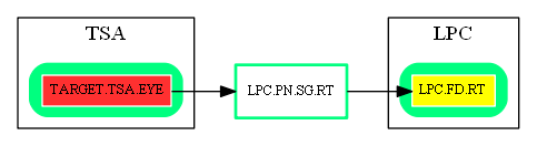
Inbound Region Connections:
| Source Area | Source Region | Source Name | Type | Reference |
| TSA | TARGET.TSA.EYE | Eye | eye-edge-output | Vision Components (EYE -> RT) |
Outbound Region Connections:
| Target Area | Target Region | Target Name | Type | Reference |
| LPC | LPC.FD.RT | Retinal Nucleus | excitatory-glu | Retinal layers (INNER -> GANGLION) |
Thirdparty Circuits
(generated)
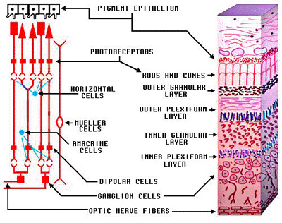
- Retinal layers - see Reference
map:
GANGLION=Retinal Nucleus,LPC.FD.RT
INNER=Retinal Ganglion,LPC.PN.SG.RT
RODSCONES=Eye,TARGET.TSA.EYE
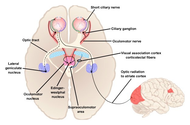
- Vision Components - see Reference
map:
CG=Ciliary Ganglion,LAC.PN.PSYM.CLG
EWN=Edinger-Westphal Nucleus,LAC.MM.EWN
EYE=Eye,TARGET.TSA.EYE
LGN=Lateral Geniculate Nucleus,NSA.FD.LGN
OCN=Oculomotor Nucleus,LPC.MM.OCN
RT=Retinal Ganglion,LPC.PN.SG.RT
SOA=Supraoculomotor Area,VBA.MM.SOA (see Reflexes Nuclei of Midbrain Mesencephalon)
STN=Spinal Trigeminal Nucleus,LPC.HL.TGS.STR
VAC=Posterior Eye Field,NPE.NC.IPS.PEF (see Intraparietal Sulcus)
VC=Occipital Lobe,NC.LOBE.OCC
References
(generated)